Questions? AskAuckland
  

LENScience


Reproductive Technologies: Genetic Screening


This page supports specialist and non-specialist teachers by providing background information about the concepts that underpin the LENScience resources on aneuploidy and related genetic screening biotechnologies.

Screening for Chromosomal Abnormalities During Pregnancy


In New Zealand, women can access pre‐natal (before birth) screening and diagnostic tests for a number of conditions including aneuploidies such as Down Syndrome. Screening is not the same as diagnosis. 


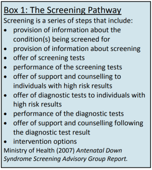
A screening test indicates the level of risk that is present for a condition such as Down Syndrome. A positive result from screening suggests that there is an increased chance of a particular condition being present and a negative result means there is a decreased risk. People with positive screening results may decide to take the next step of pre‐natal diagnostic testing which will tell them whether the condition is present or not in the fetus. Pre‐natal screening is not compulsory for anyone. Women who do choose to undergo pre‐natal screening are supported throughout the process as shown in Box 1. 


Women may decide to participate in pre‐natal screening for a number of reasons including:

  • Wanting to know as much as possible about the pregnancy 
  • Wanting to know whether or not the fetus is affected by a condition that is common in the whanau/family
  • Wanting the option of termination of an affected pregnancy 
  • Wanting to know if the pregnancy is affected by a condition so that she and her partner can prepare in advance for a baby with an abnormality


Some people prefer not to participate in screening programmes. There is a small risk of miscarriage associated with some screening procedures. A false-positive or high-risk result for an otherwise normal, healthy fetus may cause expectant parents to experience considerable stress and anxiety during pregnancy.


Conditions that can be tested for in New Zealand by pre‐natal testing include: 

  • Aneuploidy syndromes (Down [Trisomy 21], Edwards [Trisomy 18] and Patau [Trisomy 18])
  • Aneuploidy syndromes affecting the sex chromosomes (Turner [XO], Klinefelter [XXY] and Jacob [XYY]) 
  • Physical abnormalities including neural tube defects (spina bifida), cardiac, renal and digestive abnormalities
Top

Prenatal Screening


Prenatal Screening and Tests Available in New Zealand

Maternal Age

Pregnant women over the age of 35 are offered the opportunity to have diagnostic testing for chromosome abnormalities


First Trimester Screening

This involves both ultrasound Neucal Translucency (NT) and biochemical (blood test). The blood test measures hormone and protein levels which are known to indicate an increased risk of a number of abnormalities. Many pregnant women have an ultrasound scan during the first trimester of the pregnancy. The Neucal Translucency (NT) test is carried out between 11 and 13 weeks. In this scan, an increase in the fluid‐filled area at the back of the fetal neck is an indicator of an increased risk of Down Syndrome


Second Trimester Maternal Serum Screening

This uses a blood test to measure hormone levels in the mother. This information is combined with information about her age, height, weight and pregnancy dates to give an estimate of the risk of chromosomal abnormalities


Second Trimester Ultrasound Scan

This can detect many physical abnormalities, risk factors for chromosomal abnormalities as well as monitoring the growth of the fetus

Prenatal Diagnostic and Tests Available in New Zealand

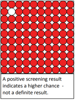
If the screening tests give a positive result, this does not mean YES or NO. It means that there is a higher chance of chromosomal abnormality. E.g. it may mean the woman is told that there is a 1 in 100 risk of Down Syndrome. If the screening result is positive, the opportunity to have diagnostic tests will be offered. These will give a definite result. Two methods of pre‐natal diagnosis are available:


1. Chorionic Villus Testing - a fine needle is inserted through the woman’s abdomen and cells are removed from the placenta. These cells are analysed in the laboratory to show chromosomal abnormalities. This test can be carried out at 11‐13 weeks and carries a 1‐3% increased risk of miscarriage. 


2. Amniocentesis - a fine needle is inserted through the woman’s abdomen and a small sample of amniotic fluid is removed. Cells from this fluid are analysed in the laboratory for chromosomal abnormalities. This test is carried out between 15 and 19 weeks and carries a 1% increased risk of miscarriage.

Top

PGD and FISH


Pre-Implantation Genetic Diagnosis (PGD)

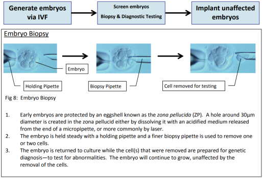
Pre‐implantation genetic diagnosis is a technique that enables couples at risk of producing embryos with genetic abnormalities the opportunity to have their embryos screened before a pregnancy is established. It is an alternative to traditional prenatal diagnosis (chorionic villus sampling or amniocentesis).


PGD can be used to screen for the sex of the embryo (in order to detect sex linked disorders such as muscular dystrophy), known single gene disorders (such as cystic fibrosis), translocations and some of the more common chromosomal abnormalities such as Down Syndrome. 


The Process of PGD 

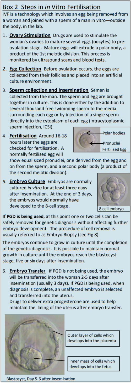
In order to carry out the genetic testing, embryos must first be formed through the use of in vitro fertilisation (IVF) as shown in Box 2. The diagnosis is performed on one or two cells removed from the embryo three to five days after conception via IVF before the embryos are implanted in the woman.  


Screening for Chromosomal Abnormalities 

As only one or two cells are available for analysis, the classical karyotype method of looking for chromosomal rearrangements is not an option in PGD as it is highly unlikely that the cells will be at a stage when the chromosomes are visible. The most common approaches use fragments of DNA tagged with fluorescent markers.


Flourescence In Situ Hybridisation (FISH)

FISH is a technique used to map the genetic material in an individual's cells, including specific genes or portions of genes. Short sequences of DNA which are chromosome specific are cloned and multiple copies made. Each specific sequence is bound to a chemical marker which will fluoresce at a specific wavelength when exposed to ultra violet light. These are commonly known as probes.


The cells obtained from the embryos are fixed onto glass slides. Fixing removes the cell cytoplasm and perforates the nuclear envelope. A solution of probes (usually limited to the eight chromosomes most commonly involved in abnormalities which can lead to failure to establish an ongoing pregnancy) is added to the slide and the mix repeatedly warmed and cooled in a special cycler.


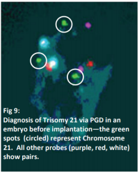
When double stranded DNA is heated up to temperatures in the range of 80 degrees celcius the two strands unbind into single stranded structures (DNA thermal denaturation). When slowly cooled the two strands recombine to form the double helix structure (DNA thermal renaturation). As the solutions contain thousands of copies of each DNA probe, there is a higher chance that a probe fragment will recombine with its complementary sequence on the test DNA than will the single copy of that chromosomes own DNA. The slide is then thoroughly washed to remove any unbound probe and studied using a microscope equipped with an ultra violet light source. A cell with the normal complement of tested somatic chromosomes will display pairs of dots of the same colour. Trisomies will show three dots of the same colour and monosomies, only one (Fig. 9).

Top

Comparative Genomic Hybridisation


The number of chromosomes able to be analysed using FISH is limited by the number of fluorescent probes which can be discriminated by the colours available. CGH allows us to look at all chromosomes using only two probe colours, usually red and green.


First the entire chromosomal complement of the cell to be studied is chopped into small fragments through the use of restriction enzymes. The fragments are copied using the Polymerase Chain Reaction (PCR). As with FISH, the DNA is heated until it becomes single stranded, but in a ‘soup’ of nucleotide elements which have been bound to a red fluorescent probe. On cooling, these labelled nucleotides anneal to the single strands to make new double stranded DNA. The process of thermal denaturation and renaturation is repeated many times until there are in order of one million copies of each fragment. During the cycling, a control cell (always from a normal male) undergoes the same process, but with green labelled nucleotides in the soup.


The test and control fragments are mixed then added to a slide on which a normal male karyotype has been fixed (the chromosomes are visible), and the slide is subjected to more sequences of warming and cooling. The fragments from the test and control genomes then competitively bind to their complementary sequences on the intact chromosomes.


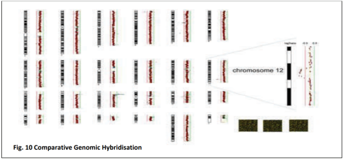
A computer aided image analyser then scans each chromosome for the presence of red or green probe. If the test genome is normal male, there will be an equal proportion of red and green probes bound throughout all chromosomes. If the test cell has an extra chromosome, for instance, a trisomy of chromosome 21 (Downs Syndrome), there will be 50% more red labelled fragments competing for sites on chromosome 21, and the chromosome on the karyotype slide will be predominantly red. Conversely, if a chromosome is absent, a monosomy, there will be 50% more control fragments of DNA from that chromosome, so it will be predominantly green. A test cell which is normal female will have a predominantly red X chromosome (as there would be two X chromosomes against the single X from the male control) and no binding on the Y chromosome.  All other somatic chromosomes will have an equal amount of red and green binding (Fig. 10)


With CGH, it is also possible to detect chromosomal deletions (parts of chromosomes which are missing) and duplications (extra copies of entire sections of chromosomes) as within the chromosomes there will be whole sections predominantly green or red respectively.


Disadvantages of CGH are either failure of the PCR process where the test and control have significantly different numbers of fragment copies before the hybridisation against the normal karyotype, and the time it takes to amplify an entire genome (several days against FISH which takes a few hours).


As the majority of chromosomal abnormalities observed in human embryos are generated at the first and second meiotic division of the oocytes (egg), the time to analysis problem can be addressed by performing CGH on polar bodies. By performing Polar Body Biopsy (essentially the same technical procedure as embryo biopsy, but the polar bodies are removed, one before insemination/injection, and the second around 18 hours later) and looking at their chromosomal complement, an inference can be made on the number of maternal chromosomes remaining in the mature and fertilised oocyte (egg).

Top

Screening for Single Gene Defects


Polymerase Chain Reaction (PCR)

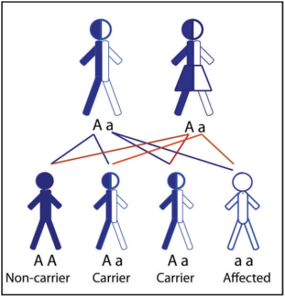
The most common technique used to detect mutations in single genes where the coding sequence of the wild type (normal) gene is known. They can be differentiated from the mutation which causes the genetic disease by size (if the mutated gene has nucleotides deleted) or by electric charge (if the mutated gene has a nucleotide substituted for another). For PCR to accurately detect the genetic inheritance of an embryo, the structures of genes of both parents must be known.


The DNA from the test cell nucleus is digested with specific restriction enzymes which will form a fragment of DNA containing the gene of interest. As with CGH, the fragments are repeatedly warmed and cooled in the presence of basic DNA building blocks until up to one million copies of each gene are present. Drops of the highly amplified DNA are placed on lanes on a gel alongside copies of the parent DNA and other controls. An electric charge is placed across the gel and the natural charge of DNA will cause the fragments to migrate. After a few hours, the distances travelled by the various fragments can be compared, therefore identifying which genes have been inherited from which parent. Unaffected or carrier embryos can then be selected for transfer. 

Pre‐Implantation Genetic Haplotyping (PGH)

In some cases the identity of the gene which causes an inheritable gene are not known, though there is often enough evidence to narrow down its location to a chromosome or part of a chromosome. Much of each chromosome consists of non‐coding DNA, i.e. they are not genes, but are inherited the same way as coding genes. As they are non‐coding, mutations are not selected against, so over generations they can become very different between individual chromosomes. There are certain regions on each chromosome which are highly polymorphic and can be used to identify which chromosome has been inherited from which parent. If the DNA of enough family members can be analysed, the chromosome bearing the otherwise unknown disease bearing gene can be identified. Rather than the test cell undergoing the PCR process to look for a gene, it undergoes PCR to identify which polymorphic markers have been inherited from each parent. The inheritance of a marker linked to the chromosome common to affected individuals is then selected against when choosing an embryo to replace.

Top

Genetic Counselling


Genetic Counselling is a communication process that deals with the human problems associated with the occurrence, or the risk of occurrence, of a genetic disorder in a family. The process involves an attempt by one or more appropriately trained persons to help the individual or the family to:

1. Comprehend the medical facts including diagnosis, course of the disorder and available management 

2. Appreciate the way heredity contributes to the disorder and the recurrence risk for specific relatives   

3. Understand the options for dealing with the recurrence risk:

  • Another pregnancy +/‐ prenatal diagnosis
  • PGD
  • Adoption
  • Artificial insemination by donor sperm or use of a donor egg
  • Childlessness 

4. Choose the course of action which seems appropriate to them in view of their risks, their family goals and act in accordance to that decision   

5. Make the best possible adjustment to the disorder and/or to the recurrence risk 

(Adapted from American Society of Human Genetics, 1975).

Top

The Human Assisted Reproductive Technologies Act (2004)

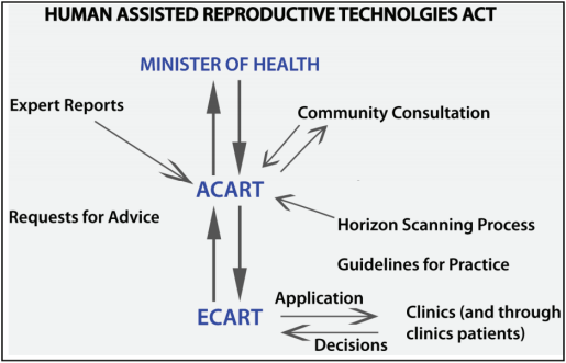
The practice of reproductive medicine in New Zealand is regulated under the Human Assisted Reproductive Technologies Act. This act established an advisory committee to the Minister of Health, called the Advisory Committee on Assisted Reproductive Technologies (ACART), whose job it is to assess information about both established and emerging new treatments and to advise the Minister of Health as to how they should be further introduced, regulated, and supervised.   


ACART is a twelve member committee representing various community interests including ethics, children’s rights, Māori interests and science. Broadly, ACART’s advice to the Minister was that a particular procedure should be an “established procedure” and that there will be no legal limitations on the use of that particular technology, or an “assisted reproductive procedure” where individual applications need to be made to the National Ethics Committee on Assisted Reproductive Technologies (ECART), who will assess the grounds on which the request is made and give their approval or not. 


The applications are made by clinics on behalf of their patients and include medical, counselling and legal reports. ECART applications are most commonly about the use surrogacy or the use of donor gametes from certain family members (i.e. son to father, father to son, relative to relative within the same generation). The HART Act comes as both an act in itself and an order in council. The order in council allows continuous change in the regulations without resorting to parliamentary debate over each and every change. 

A genetic counsellor has a Bachelor of Science with a Masters degree in genetic counselling, followed by two years of on-the-job training and education programmes prior to registration. Currently, there are only 11 genetic counsellors working in New Zealand. Common genetic referrals are from people concerned about family histories of cancer, advanced maternal age, an abnormal screening test during the pregnancy or a family history of a single gene disorder such as cystic fibrosis. Decisions that are made about the use of PGD must take in ethical considerations. This is a complex process with many varied and relevant viewpoints. Issues that are relevant for decisions about PGD include: reproductive autonomy (personal choice); autonomy of the child (under law the fetus has few rights, the moral status of the fetus is an ongoing source of debate); beneficence (doing good); non-maleficence (not doing harm) and justice (fairness, equity and the societal impact of decisions). Cultural, social and spiritual factors will play a part in decision making for people, and create variation in the way people approach these decisions.

Top