Questions? AskAuckland
  

LENScience


Understanding Breast Cancer and Biotechnology: Case Study


This case study explores the research scientists at the Liggins Institute have done into the link between increased autocrine hGH and breast cancer.

Understanding Breast Cancer


Scientists at the Liggins Institute wanted to know if increased levels of autocrine hGH were linked in some way to the development of breast cancer. To find this out they used cultured human breast cells to study the effect of differing levels of hGH on the development of breast cancer.


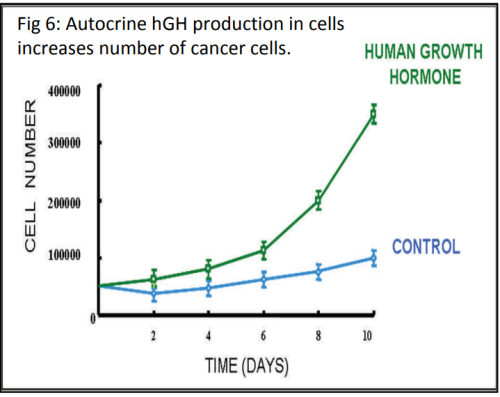
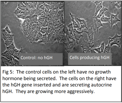
The scientists found that when hGH is being secreted from breast cancer cells, (i.e. autocrine hGH), it increases cell growth rates and the cells are more invasive (Fig 5 and 6).


Next, the scientists wanted to find out about the molecules that are regulated by hGH that were causing the cells to grow faster and more aggressively. Their suspicion was that the autocrine hGH was affecting the expression of genes in the breast cancer cells.

Cell Culture

You may be surprised to learn that human body cells are able to grow outside of our bodies if they are given the right conditions (e.g. specific pH, temperature and growth factors) and the nutrients they need to stay alive. It’s not as easy as it sounds as the right conditions require a controlled laboratory environment, especially to prevent infection by bacteria or fungi. Cells grown outside of the whole organism are said to be growing “in vitro”. This literally means “in glass”. (Cells inside an organism are said to be “in vivo” or “in life”). 


Human cells, including cancer cells, can be cultured outside the human body in plastic flasks kept in incubators. Cells grown this way are called a cell culture. Cell cultures allow scientists to test the effect of chemicals on the cells and to investigate the normal physiology or biochemistry of cells. There are many genes that scientists have noted are expressed differently in cancer cells. 


Once a gene has been identified it can be introduced to a cell line and the subsequent effects on the cells studied by growing these cells in cell culture. The gene must first be cloned and inserted into a plasmid vector and then amplified in bacteria.  The vector is then transferred into mammalian cells using cell transfection technologies such as liposomes. These cells are incubated at human body temperatures and supplied with nutrients. By comparing the gene expression in these cells with control cells scientists can determine the changes in the cells that have been caused by the inserted gene. An alternative approach to look at the effects of a single gene is to inhibit the function of the gene or protein if the cells already express it.

1
2
Top

Gene Expression: What is Going on to Create This Phenotype?


3

When studying disease, scientists want to find out how gene expression is being affected in the cells (e.g. genes being switched on/off or increasing/decreasing the level of gene expression). The phenotypic response that we see in organisms, such as the development of breast cancer, is almost always the result of a group of genes being expressed together, rather than just one gene. Scientists have been able to study gene expression one gene at a time for many years using a biotechnological process called the polymerase chain reaction (PCR). PCR is used to amplify (make multiple copies of) DNA.


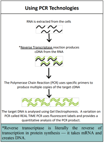
In gene expression studies, PCR is used to amplify specific genes to find out whether that gene is being expressed in the tissue being studied. If genes are being expressed then mRNA will be produced. This mRNA is extracted from the cells and a process called reverse transcriptase is used to make a short section of DNA that is complementary to the mRNA - called cDNA. The cDNA is slightly different from the original DNA because it does not contain introns (a segment of a DNA or RNA molecule which does not code for proteins and interrupts the sequence of genes). The amount of cDNA produced will depend on the amount of mRNA which is determined by how active the gene is.


The cDNA is then amplified and analysed to establish how much mRNA was in the original sample. This gives an indication of how active the gene was. PCR only allows the study of one gene at a time, which makes the process very slow. The development of a new biotechnology, microarrays, has had a major impact on research into gene expression because it allows scientists to study thousands of genes all at the same time – giving the ability to study a genetic profile. Using a microarray we can identify which specific genes a cell is using at a particular point in time. This means that we can compare which genes are turned on or off in different conditions (e.g. when cancer is present compared to when cancer is absent). 

What is a Microarray?

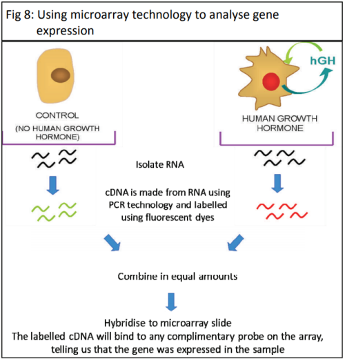
A microarray is a small glass slide that contains tiny fragments of known DNA sequences in different spots on a slide. This is also known as a gene or DNA chip. A human genome microarray will contain small fragments of each of the genes in the genome. These are called probes. Each spot on the slide contains multiple copies of the same probe. 


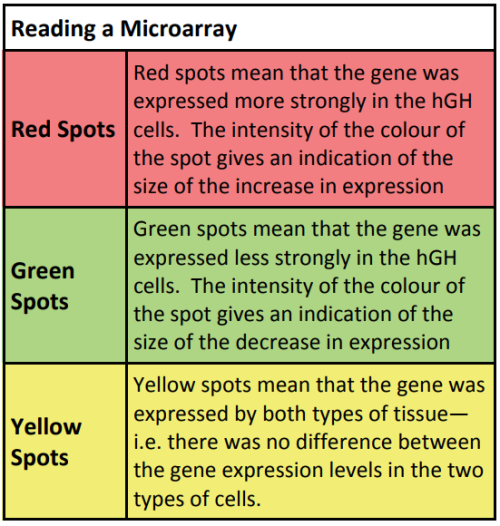
First, cDNA is extracted from the cultured cells and fluorescently labelled. When the labeled cDNA solution is washed over the slide, the fluorescently labeled cDNA pieces that match the complimentary base pairs on the slide will bond. When the slide is washed, the bonded cDNA fragments will remain in place and the other fragments will wash away. The fluorescent spots on the slide are read are read using a microarray scanner and the levels of fluorescence intensity analysed with specialised software. 

Top

The Experimental Model


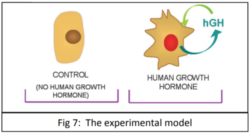
The scientists suspected that the hGH produced and secreted by the breast cells was changing the expression of genes in the cells that controlled the cell cycle. To test this the scientists created two cell lines. One contained the gene for hGH and the other (the control) produced no hGH (Fig 7).


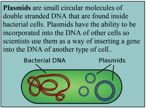
To get the gene into the cells the scientists used biotechnological techniques. First, the hGH gene was isolated and multiple copies made using PCR. The hGH gene fragments were then inserted into a bacterial plasmid. The plasmids were inserted back into bacterial cells and the bacteria grown in culture. This is a quick and relatively easy way of getting large numbers of the plasmid vector. The plasmid vectors were isolated and then inserted into human breast cancer cells that are then grown in cell culture.


A second set of identical cells that did not have the hGH gene inserted are also grown in tissue culture. PCR and microarray technologies were then used to analyse and compare the gene expression between the two cell lines. (Fig 8).


4
6
5
Top

What Did the Scientists Find?


8

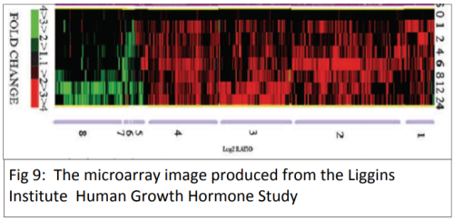
The Liggins Institute breast cancer study used a microarray analysis of 19,000 genes and found that a subset of 305 genes that were behaving remarkably differently when human growth hormone was secreted from the cells in the cell culture.


The microarray results showed which genes were turned on or off in the cells that were secreting hGH compared to the cells that were not secreting hGH. Because the role of some of the genes was known, the scientists could identify which part of the cell cycle was being disrupted. For example, if a gene that is known to play a role in making sure that damaged cells are destroyed is turned off, this helps in understanding why the cancer cells are reproducing. In addition to the genes that they knew the function of, some of the genes that were behaving differently had not been previously associated with cancer.


Scientists are now studying these genes in the hope of finding their function in the cancer cell and improving their understanding of breast cancer.

7
Top

Meeting Human Need: Personalised Treatments Derived from Genetic Information


The type of therapy used to treat a patient will depend on the type of cancer being treated and the stage of the disease.


Traditional cancer treatments include: 

  • Surgery - removal of the tumour 
  • Radiotherapy - destruction of the tumour using ionising radiation 
  • Chemotherapy - uses drugs which are effectively cellular poisons to target rapidly dividing cells or tissue that has low levels of oxygenation. Targeting rapidly dividing tissues causes problems as tissues such as the lining of the stomach are rapidly dividing and in traditional chemotherapy side affects such as an inability to hold down food and a loss of hair results from these broad target drugs


Some cancer tumours are not well supplied with blood vessels and have low levels of oxygenation - these are hypoxic. Drugs have been designed that target hypoxic tissues. 


Recent Advances in Cancer Therapy

In the last few decades there have been a number of advances in cancer therapy as a result of new technologies and improved genomic analysis of cancer. These include targeted therapies and the use of combinational therapies. In addition, individualised therapies hold great potential for the future.


Targeted Therapies

Targeted therapies are drugs that block the growth and spread of cancer by interfering with specific DNA or protein molecules involved in cancer. These therapies can be developed specifically to match the genetic and molecular characteristics of a patient’s tumour. By targeting molecular and cellular changes that are specific to cancer, targeted therapies may be more effective than conventional treatments (such as chemotherapy) and less harmful to healthy cells.


Tamoxifen was one of the first targeted therapies developed for breast cancer. The majority of breast cancers require the hormone estrogen to grow. Tamoxifen attaches to the estrogen receptor on the cell and stops estrogen from binding. When the estrogen receptors in the cell are blocked, the cell dies.


Another example of a targeted therapy used in breast cancer is Herceptin. 25% of breast cancers have a high level of the Her2 receptor which leads to increased cell growth. Herceptin is an antibody specific for Her2 which blocks the function of this receptor by binding to it. This means that the cell is no longer getting the stimulus to divide and grow rapidly and will stop growing and most likely die.


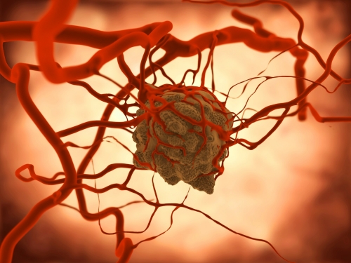
Tumour iStock_000020400324Medium

Combined Approaches to Cancer Therapy

Depending on the type and stage of the cancer, combined therapy (which uses more than one treatment) can be more advantageous than using a single agent alone. This is due to a number of reasons. Some drugs may enhance the effectiveness of another when used in combination whereas some treatments may be more effective at different stages of cancer progression. In addition, the tumour may contain several sub‐populations of cells that are very similar, but not identical. Consequently, a single agent may not wipe out all the cancer cells, leaving some to repopulate the tumour. A combination of agents has more chance of killing all the tumour cells.


Individualised Therapy

No two cancers are exactly the same. By using molecular profiling doctors can identify those patients which are unlikely to benefit from a particular therapy, or who may suffer severe side effects from a particular treatment.


This can be achieved using technologies such as microarray and real-time PCR. This development has come about through advances in technology, in particular, the human genome project, which has allowed scientists to look at genetic differences between individuals. It allows therapies to be tailored to an individual’s needs.


Using the example of Herceptin, if we give a patient with Her2‐positive breast cancer Herceptin it has the potential to be an effective treatment. If we give Herceptin to a breast cancer patient who does not have high levels of the Her2 receptor, it will most likely have no affect at all. Potentially, by using microarray analysis, multiple genes can be analysed at once leading to improvements in the choice of therapies that are used.


Using Biotechnologies to Understand Cancer

In order to treat cancer effectively, scientists need to know as much as possible about what is causing the cancer and the way the cancerous tissue behaves in different environments. They want to find out what increases the growth rate of the cancers and what can reduce the growth. This knowledge will improve our ability to find effective treatments for cancers. Scientists at the Liggins Institute in Auckland are investigating the role of growth hormone in breast cancer. Biotechnologies play a major role in this investigation and the consequent development of potential therapies.


Technology

How This Technology Has Helped Scientists' Understanding of Cancer

Gene Profiling About 5‐10% of cancer patients inherit a genetic defect that gives them a susceptibility to cancer over their lifetimes. Many years of research using a number of biotechnologies went into achieving this understanding for example: PCR, DNA sequencing and gene mapping. Gene profiling allows scientists to identify individuals that carry specific alleles that increase their risk of developing cancer in their lifetime.
Genome Analysis

Scientists have been able to study gene expression one gene at a time for many years using PCR technology. Primers specific to the gene of interest were used in the PCR mix to find out whether that gene was being expressed in the tissue.   


Microarrays are used to compare the expression levels of thousands of genes all at the same time, enabling scientists to study a genetic profile. The microarray technology can identify which specific genes a cell is using at a particular point in time. This means that we can compare which genes are turned on or off in different conditions (e.g. when cancer is present compared to when cancer is absent).   
 

The information from the microarray gives an overview of which genes are turned on or off, over or under expressed. To confirm how much more or less a gene is being expressed in a cancer cell line “Real-Time PCR technology” is used. This is using PCR with primers specific to the gene as usual but a real time PCR allows scientists to further quantify the change in gene expression.       


Often the microarray analysis will identify genes that are already known to have a role in cancer. However, sometimes an experiment will identify a known gene which has not previously been associated with cancer. This is useful information as it identifies new targets for cancer therapy.

Top

References


Mukhina, S.,  Mertani, H C., Guo, K., Lee, K. O., Gluckman,  P. D., & Lobie, P. E. (2004). Phenotypic conversion of human mammary carcinoma cells by autocrine human growth hormone. Proc Natl Acad Sci U S A 101(42), 15166‐15171. https://doi.org/10.1073/pnas.0405881101


Xu, X. Q., Emerald, B. S., Goh, E. L., Kannan, N., Miller, L. D., Gluckman, P. D., … Lobie, P. E. (2005). Gene expression profiling to identify oncogenic determinants of autocrine human growth hormone in human mammary carcinoma.  J Biol Chem 280(25),  23987‐24003. https://doi.org/10.1074/jbc.M503869200

Top