Questions? AskAuckland
  

LENScience


What is Breast Cancer?


This page supports specialist and non-specialist teachers by providing background information about the concepts that underpin the LENScience resources on breast cancer and biotechnology.

Breast Cancer


feature

Each year in New Zealand, approximately 3300 women and 25 men are diagnosed with breast cancer and approximately 600 people die of breast cancer. 70% of those diagnosed will be over the age of 50 years. Over the last 10 years, the number of people diagnosed with breast cancer has increased, however, very recently we have been seeing a decline in incidence in many countries which is most likely due to identification of Hormone Replacement Treatment as a risk factor. In 1960, around 1 in 30 NZ women were diagnosed with breast cancer in their lifetime; today that figure is 1 in 9. This is partly due to improvements in screening and diagnosis but breast cancer rates were also increasing. A woman in NZ has an 11% chance of being affected by breast cancer.


While the number of women being diagnosed with breast cancer has been increasing, the good news is that the death rate from breast cancer decreased by around 27% in the past decade. This is a result of increased understanding of the disease, improved screening, diagnostic methods and treatment. New Zealand has a large and active cancer research community that plays an important and often world-leading role in cancer research. Biotechnology has played a significant role in the development of knowledge of cancer and treatment of this disease.

Top

What is Cancer?


1

Healthy cells have a fixed life span. They replicate (reproduce) a certain number of times and then enter a period of senescence where they stop dividing. This is followed later by cell death (apoptosis). Cancer occurs when cells keep growing instead of dying. This causes the development of tumours which disrupt normal body function. Cancer may occur in any part of the body. Tumours may be solid masses of cells such as a breast cancer tumour, or they may be liquid tumours consisting of blood cells that have become cancerous such as in lymphoma and leukaemia.


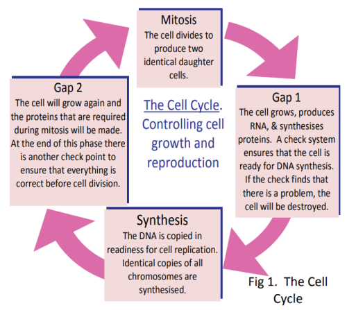
The cell cycle (Fig 1) controls normal growth and reproduction in cells. It is controlled by two families of molecules. Faults during the cell cycle such as inadequate growth and mistakes in the replication of the chromosomes are generally picked up by these control agents. The cell will then be repaired, or stop dividing and die later or the cell will self-destruct (apoptosis). The process of cell growth and division is regulated by signals from inside and outside the cell.


Unlike normal cells, cancer cells are immortal. They do not respond correctly to the regulatory signals and will go on replicating themselves over and over again. This fast rate of reproduction creates a mass of excess cells, called a tumour.


What Makes a Cell Become Cancerous?

Cancers arise from changes in the cell that give the cell a growth advantage. This means the cell has an increased ability to grow faster and more aggressively. The changes may be in the DNA sequence (mutations) or they may be epigenetic changes such as DNA methylation, that alter gene expression without altering the sequence of base pairs in the gene. DNA methylation can be caused by changes in hormone exposure, or exposure to chemicals in the environment and in diet. 


For more information about epigenetic changes, see the LENScience resources on gene expression.


Cancers that start with a mutation are usually a result of exposure to an environmental factor (a mutagen) but they can also be a result of errors in everyday cellular process e.g. DNA replication errors. They can also occur spontaneously. In most cases, the mutations are caught in the checks that are a part of the cell cycle and either the error is corrected or the cell is destroyed. If the cell cycle is not carefully controlled and checked, the mutated cell will survive and produce more mutated cells. Cancer is usually the result of an accumulation of mutations or changes in gene expression that transform the normal cell into a cancerous cell. As we get older, we accumulate more mutations and changes in the DNA packaging, therefore increasing the chances of cancer occurring as we age.

Top

The Hallmarks of Cancer


2

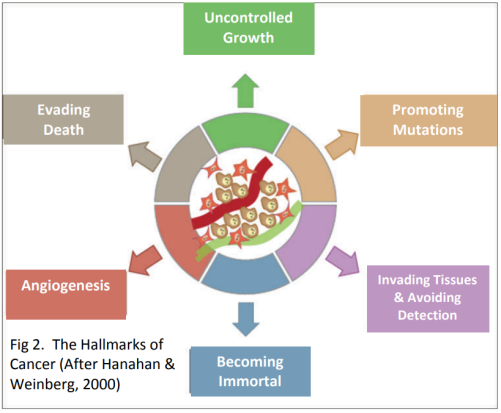
Scientists Douglas Hanahan and Robert Weinberg think that all the different types of cancers share six essential changes in the physiology of the cell. These changes all contribute to the production of a successful tumour. They called these six essential characteristics the Hallmarks of Cancer (Fig. 2).


1. Uncontrolled Growth

Normal cells start or stop growing in response to signals. Cancer cells do not respond to these signals. They will grow when there are no growth signals and continue to grow when they are getting stop signals. So no matter what colour the traffic lights are, cancer cells will go! 


2. Evading Death

Normal cells are meant to die when they get old or damaged. Signals in the cell start a process where the cell membrane is disrupted and enzymes break down the cell contents, which are then engulfed by neighbouring cells. This is called programmed cell death or apoptosis. Cancer cells avoid programmed cell death. They have the ability to repress (ignore) the signals that start the process of apoptosis.


3. Angiogenesis

This means that all cells must be in close proximity (within 100µm) of a capillary if they are to survive. Growth of a tumour is not possible past a certain size unless the cells are capable of attracting blood vessels to grow into the tumour in order to supply nutrients and remove wastes.   We say a tumour is angiogenic if it has the ability to attract blood vessels to grow into it.


4. Becoming Immortal

Normal cells have a limited number of times that they can divide before they stop growth. Sequences of DNA on the ends of chromosomes called telomeres are responsible for making sure that chromosomes do not fuse end‐to‐end during cell division. Each time a cell divides, the telomeric DNA gets slightly shorter. When it gets so short that it cannot protect the chromosome anymore, the cell dies. In cancer cells the telomeric DNA does not get shorter because an enzyme called telomerase is released that lengthens the telomeres so the cell can keep on replicating endlessly. 


5. Invading Other Tissues and Avoiding Detection (Metastasis)

It is common for cancers to move from the site of the primary tumour to other parts of the body.   This is called tissue invasion or metastasis. The immune system is constantly on the lookout for cells that are foreign or do not belong. Cancer cells often look different to normal cells and when spreading out will invade tissues different to their own. In order to survive, pre‐cancer cells and cancer cells must evade detection by the immune system.


6. Promoting Mutations

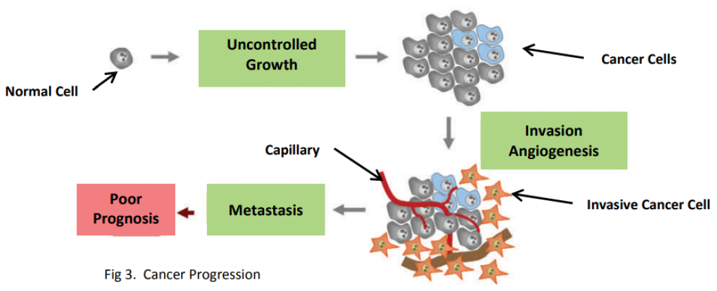
The development of cancer requires an accumulation of mutations in a number of genes over a period of time. Some of these changes accelerate the rate at which mutations occur—meaning that mutations are acquired at a faster rate. Together all these characteristics contribute to the progression of normal cells into cancer (Fig 3).


 

3

Cancer Risk Factors

Growing older Tobacco
Sunlight Ionising radiation
Certain chemicals and other substances Some viruses and bacteria
Certain hormones Family history of cancer
Alcohol Poor diet, lack of exercise or being overweight
Top

Breast Cancer and Human Growth Hormone


Human growth hormone (hGH) is made and secreted by the pituitary gland, which is located at the base of the brain. Human growth hormone is essential for normal growth and development in humans. Not enough hGH in childhood results in a person who is very short while excess growth hormone results in gigantism.


In addition to the hGH that is produced and secreted from the pituitary gland (which is part of the endocrine system), hGH is also produced and secreted locally in different sites around the body. (Locally means the hormone affects only that area). These sites include parts of the central nervous system, cells within the immune system, blood vessels and breast tissue. 


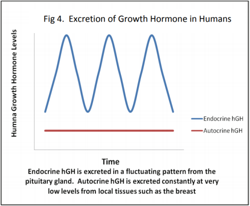
The levels of hGH secreted from the pituitary gland fluctuates during the day. It has high peaks and low troughs. In contrast, the hGH that is secreted from the local tissue sites, such as breast tissue, is secreted continuously at very low levels (Fig 4).


Studies conducted at Liggins Institute at the University of Auckland have demonstrated that hGH secreted locally in the breast tissue plays a critical role in the development of breast cancer. Autocrine hGH secreted in the breast is essential for normal breast development during puberty. However, Liggins scientists have found that there are increased levels of autocrine hGH found in breast cancer tumours. Read the case study resources to find out more about their findings and developments in cancer treatments.


Endocrine Hormones that are secreted into the bloodstream and carried to the target tissues or cells
Autocrine Hormones that only affect the type of cell that produced them
4
Top

References


Breast Cancer Foundation NZ. (2019). Breast cancer. Retrieved from https://www.breastcancerfoundation.org.nz/breast-cancer


Hanahan, D., & Weinberg, R. A. (2000).  The hallmarks of cancer. Cell, 100, 57‐70. https://doi.org/10.1016/S0092-8674(00)81683-9


Perry, J. K., Emerald, B. S.,  Mertani, H. C., & Lobie, P. E. (2006). The oncogenic potential of growth hormone. Growth Hormone & IGF Research, 16, 277–289. https://doi.org/10.1016/j.ghir.2006.09.006

Top