Questions? AskAuckland
  

LENScience


Understanding Huntington's Disease: Case Study


This page presents more about the work of scientists at Auckland University and the Centre for Brain Research in the prevention and treatment of Huntington’s disease.

Living with Huntington's Disease


A number of people with HD and their families have shared their stories using YouTube. In sharing their experience they hope to encourage people to understand more about the disease and why finding a cure is so important.


NHS - Choices: Huntington’s Disease (Lee, Aged 39)

Lee’s mother had Huntington’s disease and Lee himself is affected. Lee talks about life with Huntington's disease.


CBS Special - Huntington's Disease

Watch to learn more about the impact of Huntington’s on individuals and families.


Treating Huntington’s Patients

The Hartwyk Centre is a care facility in New Jersey that cares for Huntington’s patients.

Top

Animal Models


81295672

By studying donated brains from the Brain Bank, Professor Faull’s team have shown that neurons containing γ-aminobutyric acid (GABA), a neurotransmitter that acts as a signalling chemical between neurones, are selectively lost in the striatum of the basal ganglia of humans with HD. Unfortunately, it is not possible to treat patients with GABA because of the effect this treatment would have on the rest of the brain. 


While investigating the disease in humans is crucial, the use of animal models has also become an important tool for discovering information about many aspects of HD. These animals (e.g. transgenic mice, fruit flies and nematodes) can be manipulated far more easily than humans, progress more quickly to show the symptoms of HD and have shorter life-spans; therefore these models have a huge advantage over the study of humans as it can speed up the rate of research and information gathering. In addition, the brains of these animals can be accessed early, rather than having to wait until death occurs naturally. Animal models are being used to search for potential drug treatments, with a hope that the outcomes can be translatable to the human disease. 


Professor Faull’s team use rats and induce HD by injecting the toxin quinolinic acid into the basal ganglia region of the brain. This toxin is present at higher than normal concentrations in the brains of humans with HD and induces similar symptoms in rats. In addition, the team have transplanted basal ganglia brain cells from a fetal rat into diseased adult rats and found that these cells survive and make new non-diseased GABA-like brain cells. There is hope that the transplantation of fetal brain cells may one day help humans with HD. A similar treatment for Parkinson’s disease has already been trialled in which cells producing the required neurotransmitter dopamine were transferred. Unfortunately the treatment was only partially successful as it resulted in tumour formation in some cases. 


While some discoveries in animal models can be directly applied to humans, scientists are always very aware that in many cases this is not possible. Tissue culture studies are another tool to extend our understanding. The cells in tissue culture are isolated – rather than in whole organisms – and investigating these cells in isolation can reduce the ‘noise’ associated with studying the full organism. However, these cells may behave differently when they are isolated from the whole organism, therefore information gathered from isolated cells needs to be further investigated with a view to the whole organism. 


While no one tool offers all the answers, it must be kept in mind that all these tools play an important part in extending our understanding of the disease and will eventually enable scientists to finding a cure for HD.

Top

The Brain's Regenerative Potential


Scientists have now disproved the long-held belief that humans are born with all the brain cells they will ever have. They now know that the diseased adult human brain tries to repair itself by making new replacement brain cells, in a process is called neurogenesis. 


In 2003, University of Auckland scientists published a paper showing that the brains of HD patients had been trying to regenerate. Using the Brain Bank, they took sections of normal and HD brains, and showed that there was more cell proliferation and growth in the diseased brains than in the normal brains. Their current hypothesis is that the markers of cell proliferation and growth that they discovered are due to the brain trying to grow new cells to compensate for the destroyed ones. They are hopeful that it might one day be possible to treat brain diseases like HD by augmenting this normal response.

Top

Stem Cells: A Hope


2

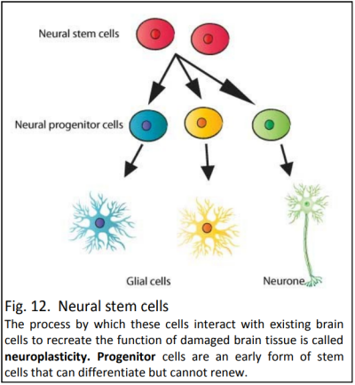
The cells responsible for the cell proliferation and growth found in adult brains (and more so in HD brains) are neural stem cells, a form of an adult stem cell. 


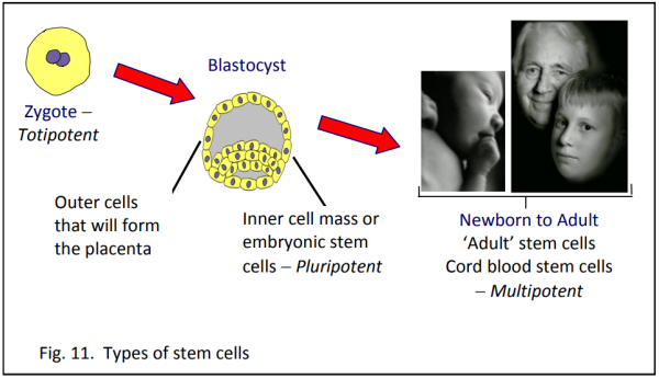
Stem cells differ from other cells in that when they go through cell division they can produce undifferentiated cells or, under the right conditions, they have the potential to differentiate to form specialised cells, with a specific function, e.g. muscle cells, skin cells, nerve cells. It is important to distinguish between embryonic stem cells and adult stem cells (Fig. 11).


Embryonic Stem Cells

Every cell in a zygote – the group of cells that forms soon after fertilisation – is totipotent, having the ability to form all types of cells. 


Embryonic Germ Cells 

The inner cells in a blastocyst – the next step on from a zygote – are pluripotent, having the ability to form all cell types in the body except the placenta (the outer cells form the placenta). 


Adult Stem Cells 

These are undifferentiated cells that are yet to specialise and are found in infants, children and adults. They can only produce a limited number of cell types specific to the tissue they’re found in, and are called multipotent. Tissues that have been found to contain adult stem cells include the brain, bone marrow, skeletal muscle and skin.


Embryonic stem cells, therefore, have the most flexibility. However, they are associated with major ethical challenges, because of the sources of these cells - either an embryo that is excess from IVF (which is very controversial) or more simply they can be extracted from cord blood when a child is born. 


1

In theory adult stem cells can be extracted from an adult, grown and replaced into the same person, which avoids the ethical problems of embryonic cells. Replacing stem cells into the person they came from also avoids problems with rejection by the immune system when it recognises foreign cells, an issue that would be present in embryonic stem cell therapy other than from cord blood. 


All stem cells have a ‘homing’ ability, enabling them to migrate to the area where new cells are needed. In 2007, a paper published in one of the world’s most prestigious journals, Science, first showed the route that progenitor cells stream through in the human brain. This research was led by University of Auckland scientists. Most stem cell lines grown in labs around the world for the purposes of research are embryonic stem cells. Scientists from the Centre for Brain Research are growing stem cells cultivated from brains donated to the human brain bank, both normal and diseased. They are using them to examine things such as whether adult stem cells from HD brains are different from those in the non-diseased brains and whether they have the potential to form significant numbers of “good” new brain cells to slow (or even halt) the process of brain cell degeneration. 


The potential to replace destroyed brain cells using stem cells is very exciting, but there are still many challenges to overcome. Researchers need to discover the precise environment (chemicals, growth factors and more) which will successfully direct the adult stem cells to make just the right type of new brain cells in the right numbers and in the right region of the brain. In particular, one of the dangers of stem cell therapy is the potential for stem cells to multiply uncontrollably, leading to tumours that cause more damage than the original disease. 

Top